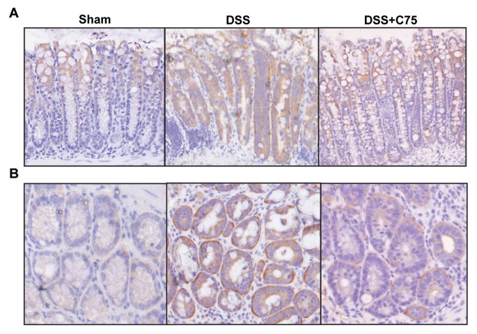
Figure 3.
Expression of FASN in the colon of DSS colitis. Colon tissues from sham, vehicle and C75-treated groups at d 8 were subjected to immunohistochemical analysis against FASN with brown staining and counterstained with hematoxylin. Representative images of longitudinal sections (A) (100× magnification) and transverse sections (B) (200× magnification) are shown.

