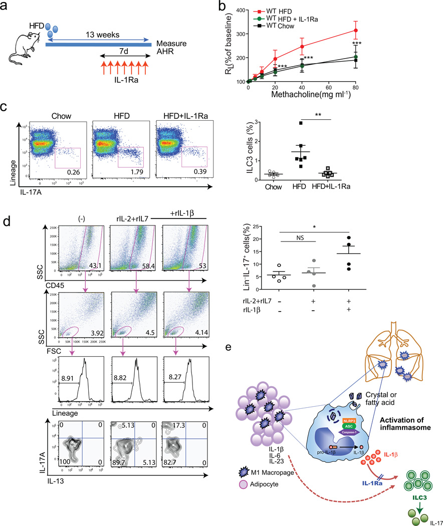
Figure 6. Treatment of obese mice with anti-IL-1, prevents AHR.
a. Schedule of feeding and anti-IL1R antagonist treatment. To block IL-1β (and IL-1α), obese mice were treated with IL1R antagonist (50 mg/kg) subcutaneously for 7 days before measuring AHR.
b. AHR was measured 24hr after last IL1R antagonist treatment. Graph represents the changes in lung resistance (RL). ***p<0.001, IL1R antagonist treated obese mice were compared to HFD fed obese mice. (Two-way ANOVA).
c. ILC3 cells were analyzed after PMA/Ionomycin stimulation (left panel). Graph represents the percentage of IL-17A producing ILC3 cells of the total lung lymphocytes in each group (right panel).
d. Cells from BAL fluid were cultured in vitro in the presence of rIL-2, rIL-7 and rIL-1β for 72hr. To measure IL-17 production from Lin− population, cells were re-stimulated with PMA and ionomycin for 5hr. Graph represents the percentage of IL-17 producing CD45+Lin− lymphocytes in each group (bottom), *p<0.05 (Student’s T-test).
e. Possible mechanisms of obesity induced AHR. High fat diet results in the activation of the NLRP3 inflammasome, through fatty acids or cholesterol crystals in macrophages in adipose tissue and in the lungs. This results in IL-1β production, which drives the development of ILC3 cells in the lungs. Treatment with IL-1R antagonist blocks the effects of IL-1β (and IL-1α), and blocks the development of pulmonary ILC3 cells.

