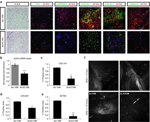
Figure 4.
ALK5ViM treatment of Dupuytren's disease (DD) resection specimens cultured in the three-dimensional (3D) ex vivo “clinical trial” system. (a) Immunohistochemical and immunofluorescent analysis of 3D cultured DD resection specimens after 3-day treatment with scrambled ViM (ScrViM) and ALK5ViM. Hematoxylin and eosin (H&E), proliferation marker phosphohistone 3 (PH3, green), apoptosis marker cleaved caspase 3 (CASPASE3, green), collagen type 1 (COL1A1, green), collagen type 3 (COL3A1, green), phosphorylated SMAD2 (pSMAD2) (green) and smooth muscle actin, α2 (ACTA2, red). Nuclei were visualized with TOPRO-3 (TOPRO, blue). (b) Quantitative polymerase chain reaction (Q-PCR) to detect expression levels of full length ALK5 mRNA was performed on tissues injected with ScrViM and ALK5ViM (N = 3). Values were normalized to CAPNS1. Fold induction values compared to ScrViM condition are shown. Statistical significance was calculated by one-tailed paired t-test. ***P < 0.001. (c–e) Quantification (described in methods) of immunofluorescence signal of COL1A1, COL3A1, and ACTA2 from different patient-derived specimens (N = 3) after 3-day treatment with scrambled ViM (ScrViM) and ALK5ViM. Fold induction values compared to ScrViM condition are shown. Multiple areas were quantified per sample, error bars represent ± SEM. Statistical significance was calculated by one-tailed paired t-test. *P < 0.05, **P < 0.01. Scale bars, 25 µm. (f) Second harmonic generation (SHG) images of endogenous DD tissue in the 3D culture system. Collagen distribution was imaged at control time point (SHG, t = 0, upper panel) in adjacent parts of the specimen. The exact tissue parts were imaged at 72 hours (SHG, t = 72 hours, bottom panel) after injection of ScrViM or ALK5ViM. Arrow indicates site of injection.

