Figure 2.

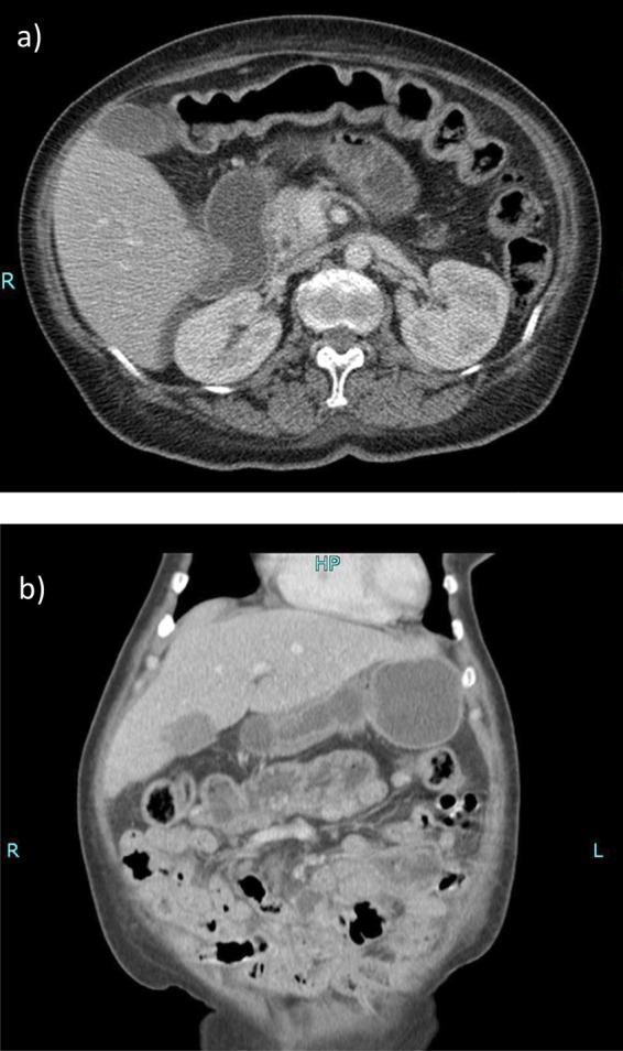
Contrast-enhanced CT scan of the abdomen: prominent fluid-filled loops of small bowel are seen. The duodenum and proximal small bowel are moderately distended and fluid filled. Axial section at the L2 level (A). Coronal section (B).

Contrast-enhanced CT scan of the abdomen: prominent fluid-filled loops of small bowel are seen. The duodenum and proximal small bowel are moderately distended and fluid filled. Axial section at the L2 level (A). Coronal section (B).