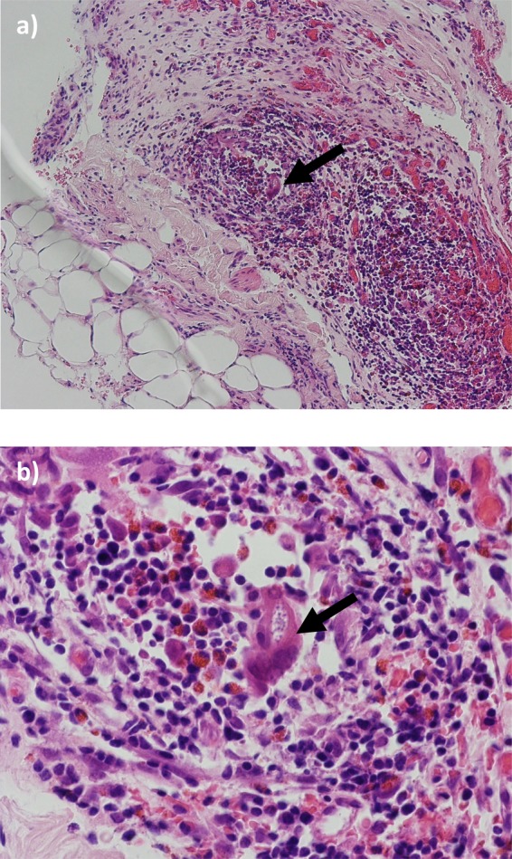
Figure 3.

H&E staining of small bowel mesentery: fibroadipose tissue with inflammatory cell infiltrate composed of lymphocytes, eosinophils and occasional plasma cells are noted. The foreign body granuloma amidst the inflammation with a prominent eosinophilic cell infiltrate is suggestive of worm infestation. (A) ×40 magnification. (B) ×100 magnification. The arrow points to the foreign body granuloma in each slide.
