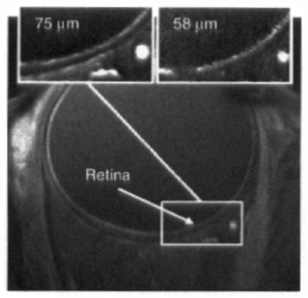
Figure 4. Representative fine resolution of human eye (courtesy of Drs. S. Lalith Talagala, Daniel Reich and Alan Korestsky (NIH) who also provided much of the following summary).
MRI was performed on a 3.0 T scanner (GE Signa Excite HDx, Waukesha, WI, USA) using a receive-only surface coil (diameter of 40 mm) connected to a head support. The coil was positioned over the right eye, without touching the face. The left eye was patched. The head was slightly tilted to the contra-lateral side. Participants were asked to fixate when necessary on a red tape X positioned on the upper internal surface of the scanner bore. Images were collected using a 2D fast spoiled gradient echo sequence (TR = 16 ms; TE = 3.4 ms; flip angle 25; matrix size 512 × 128; FOV 3.8 × 3.8 cm; slice thickness 2.5 mm; pixel size 75 × 75 μm). 20 separate images, each taking 12 s to acquire while the patient does not blink followed by 5 s of interval where rest and blinking are encouraged, were acquired. These 20 images were acquired on the same location. This cued blinking protocol has been described elsewhere [93]. Following rigid image registration (and removal of images with too much movement artifact), an average image was generated as shown. Top inserts: region of interest of images collected with resolutions of 75 and 58 μm2.

