
Figure 1. Histology and immunohistochemistry of MCC.
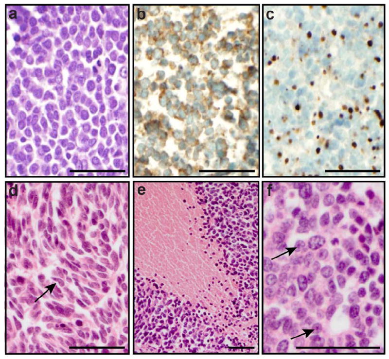
(A) H&E staining illustrating a population of monomorphic, non-cohesive round cells with a high nuclear to cytoplasmic ratio and finely granular “salt and pepper” chromatin. (B) Positive staining for the neuroendocrine marker synaptophysin. (C) cytokeratin 20 dot-like perinuclear staining. Additional features observed in some tumors: (D) spindle morphology (arrows), (E) large zone of necrosis, and (F) prominent nucleoli (arrows). Scale bar: 50 micron.