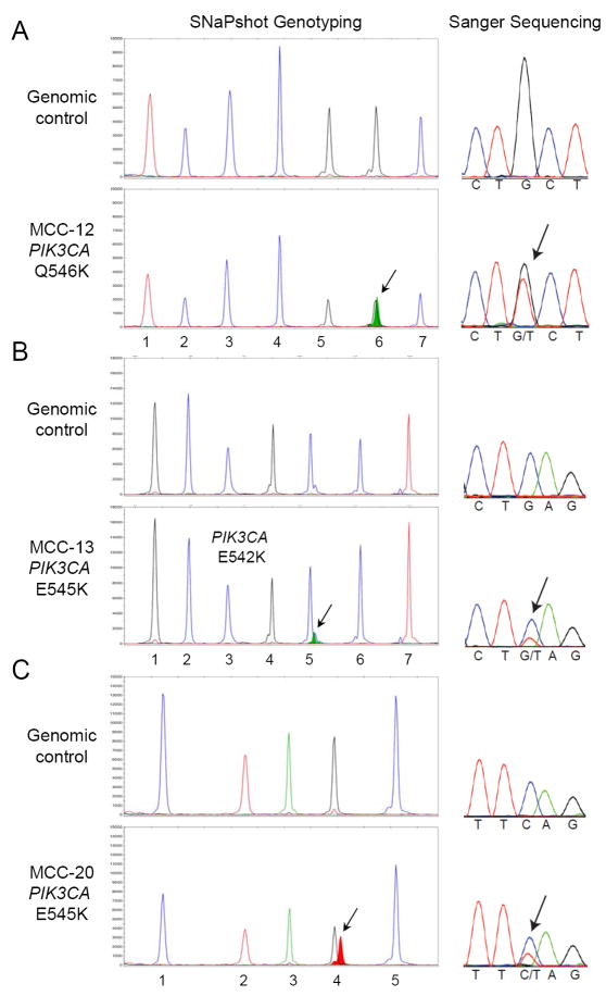
Figure 3. Detection of PIK3CA activating mutations in MCC.
Mutational profiling was performed by SNaPshot analysis (left panels) and confirmed by Sanger sequencing (right panels). Nucleic acid extracted from each MCC archival specimen was run in parallel with a genomic DNA control, as indicated. The arrows point to the mutant alleles: PIK3CA Q546K (c.1636C>A) in tumor MCC-12 (A), PIK3CA E545K (c.1633G>A) in MCC-13 (B), and PIK3CA E542K (c.1624G>A) for MCC-20 (C). The assayed loci for each panel are as follows: (A) 1.PIK3CA 3140, 2.CTNNB1 101, 3.BRAF 1798, 4.NRAS 37, 5.PIK3CA 1636, 6.APC 4348 and 7.APC 3340; (B) 1.KRAS 34, 2.EGFR 2235_49del F, 3.EGFR 2369, 4.NRAS 181, 5.PIK3CA 1633, 6.CTNNB1 94 and 7.CTNNB1 121; and (C) 1.EGFR 2236_50del F, 2.EGFR 2573, 3.CTNNB1 133, 4.PIK3CA 1624 and 5.NRAS 35.

