
Figure 4. Sensitivity of MCC cell lines to PI3K pathway inhibitors.
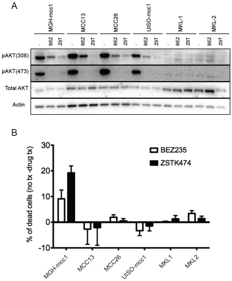
(A) PI3K pathway activation in MCC cell lines. MGH-mcc1 (PIK3CA-mutant), MCC13, MCC26, UISO-mcc1 (MCPyV+), MKL-1 (MCPyV+), and MKL-2 (MCPyV+) cells were treated with vehicle control (-), with 200nM of the dual PI3K/mTORC inhibitor, NVP-BEZ235, or with 1mM of the pure PI3K inhibitor, ZSTK474, for 6 hours. Lysates were prepared and subject to immunoblotting with the indicated antibodies. Blots were stripped and probed with α-Actin to ensure proper loading. (B) Targeted PI3K and PI3K-mTOR inhibition reduced cell viability in PIK3CA-mutant MCC. The cell lines were treated as described above for 72 hours. The cells were then stained with propidium iodide (PI) and Annexin V and analyzed by FACS. Because we found that plasma membranes of healthy MKL-1 and MKL-2 cells displayed constitutive binding to Annexin V, these cells were instead permeabilized and stained with only propidium iodide, and sub-G1 cells were quantified and scored as dead by FACS, as previously described (31).