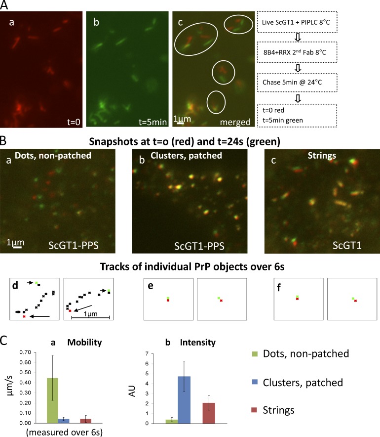
Figure 5.
Live staining: FL PrPSc strings are slowly mobile, resembling Ab-clustered PrPC patches. (A) ScGT1 were treated with PIPLC, labeled with 8B4/secondary Fab (all at 8°C), and followed by time-lapse microscopy at 24°C. Snapshots at t = 0 (red) and t = 5 min (green) are superimposed in c. (B) ScGT1-PPS (a and b) and ScGT1 (c) cells were labeled with 8B4 followed by secondary Fab at 8°C. (b) PrPC was patched with a tertiary Ab. Cells were then imaged every 400 ms for 24 s at 24°C. Snapshots at t = 0 s (red) and t = 24 s (green) are superimposed. Three types of PrP objects were followed: small dots in cells not patched with the tertiary Ab (a, uninfected), large Ab-induced clusters (b, uninfected), and strings (c, infected). (d–f) Tracks of individual PrP objects over 6 s. Starting point in red (arrow) and end point in green (arrowhead). Many small 8B4 dots (a) were too mobile to allow tracking for the 6-s period. (C) Mean mobility (a) and fluorescence intensity (b) of the three object types measured over 6 s. Both large Ab-induced clusters (blue columns) and strings (red) were mostly immobile, whereas smaller dots (green) in nonpatched samples were very mobile (∼0.4 µm/s).

