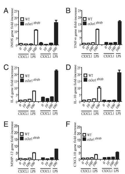
FIGURE 6.
A–F, Gene expression in interstitial lung mononuclear phagocytes obtained from cx3cr1+/+ and cx3cr1GFP/GFP mice. CD11c-enriched interstitial lung mononuclear phagocytes were harvested and pooled from cx3cr1+/+ (WT; n = 12) and cx3cr1GFP/GFP (n = 9) mice under steady-state conditions. Cells were stimulated in the presence or absence of increasing concentrations of recombinant CX3CL1 (0, 10, 100 nM) or LPS (100 ng/ml). Inducible NO synthase (iNOS), TNF-α, IL-6, IL-10, CXCL10, and MMP-12 gene expression was assayed by real-time PCR using the ΔΔCt method and 18S as the endogenous control. Unstimulated WT lung mononuclear phagocytes served as the calibrator. The y-axis represents gene fold-increase above the calibrator. Each condition was assayed in triplicate.

