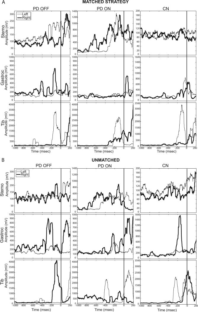
Fig 5.
Individual representative rectified EMG traces from 180° in-place turns using the matched strategy (A) and unmatched strategy (B). The left (thin line) and right (bold line) sternocleidomastoid (Sterno), medial gastrocnemius (Gastroc), and tibialis anterior (Tib) are shown from one individual with PD OFF and ON medication and one control. All turns are to the left. The vertical line at zero indicates turn onset.

