
Figure 2.
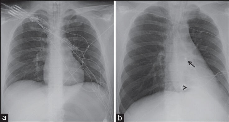
Chest radiograph obtained at admission (a) demonstrates no cardiopulmonary abnormality. A follow-up chest radiograph (b) shows a 5 mm round metallic foreign body projecting over the left hilum (open arrow) with interval development of a wedge shaped pulmonary opacity a the left lung base (arrow head)