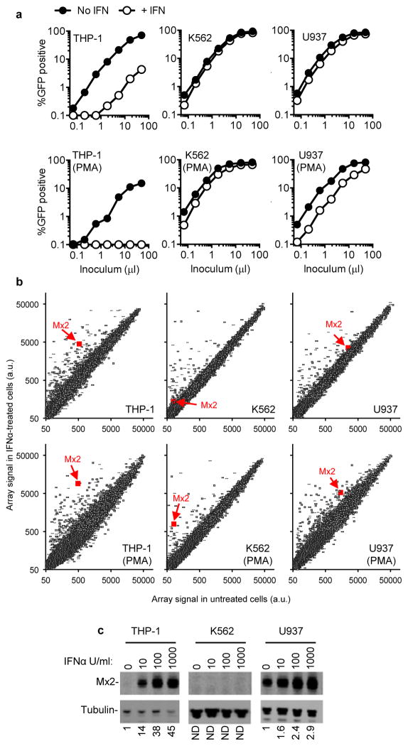
Figure 1. Differential effects of IFNα on HIV-1 infection of monocytoid cell lines correlates with Mx2 expression.
a, Undifferentiated, or PMA-differentiated THP-1, K562 and U937 cells with or without IFNα treatment (1000 U/ml) were challenged with a GFP-expressing HIV-1 vector (CSGW). b, RNA extracted from cells treated identically to those shown in a was analyzed on microarrays. The array signal is plotted in arbitrary units, and the data points representing Mx2 are highlighted. c, Western blot analysis of Mx2 and tubulin expression in monocytoid cell lines treated for 24h with the indicated doses of IFNα. Numbers below each lane indicated fold increase in Mx2 protein levels relative to untreated cells.

