
Fig. 3.
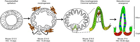
Milestones in murine and HIO intestinal development. The intestinal epithelium is present as a pseudostratified epithelium at ∼E12.5 through E13.5 in the mouse, and within 14 days of HIO specification. After this point, the appearance of mesenchymal clusters (orange), which respond to hedgehog (Hh) and platelet-derived growth factor A (Pdgfa) secreted from the epithelium, is coincident with the onset of villus morphogenesis. In the mouse, villus morphogenesis proceeds between E14.5 and E16.5, whereas in HIOs this stage takes considerably longer. As the epithelium remodels, the columnar epithelium forms with stereotypical villus (green) and proliferative intervillus (yellow) domains. Mesenchymal clusters remain associated with the villus tip and act as a postmitotic signaling center, signaling to the epithelium via secreted growth factors (including Bmp ligands, among others), which prevent epithelial proliferation. The fetal intervillus domain gives rise to the adult crypt by postnatal day 14 in the mouse, and after approximately 8 weeks in HIOs. The mature adult crypt houses the proliferative intestinal stem cells (yellow) and secretory Paneth cells (red), as well as several additional cell types, the most prevalent of which are enterocytes (green), goblet cells (blue) and enteroendocrine cells (purple).