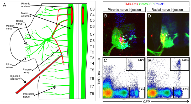
Fig. 1.
Retrograde labelling and purification of mouse E11.5 embryonic phrenic MNs by flow cytometry. (A) The phrenic nerve was injected with TMR-dextran in Hb9::GFP trunk explants. Spinal segments are numbered according to the ventral roots that emerge from them (labels on right). C, cervical; T, thoracic. (B) Mid-cervical spinal cord transverse section: phrenic neurons (arrow) are labelled with TMR-dextran (red). The Hb9::GFP transgene labels all MNs; phrenic neurons co-express Pou3f1. Scale bar: 50 μm. (C) TMR+ GFP+ phrenic neurons and GFP+ non-phrenic MNs were isolated from spinal cords (C3-C5 levels) by flow cytometry. (D) TMR+ GFP+ radial LMC neurons (arrow) in E11.5 cervical spinal cord (C6-C8 levels), following retrograde tracing through the radial nerve. (E) Purification of TMR+ GFP+ radial LMC neurons by flow cytometry.

