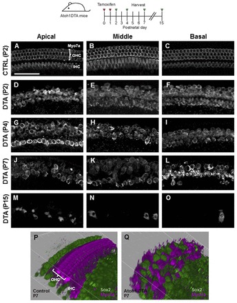
Fig. 2.

Progressive HC death in the Atoh1DTA model. (A-O) Projection images of Myo7a immunofluorescence in cochlear whole-mounts from control mice (lacking either the Cre or DTA allele) at P2 (A-C) and Atoh1DTA mice at P2 (D-F), P4 (G-I), P7 (J-L) and P15 (M-O). Repopulation of HCs was most robust in the apical turn at P4 (G). (P,Q) 3D reconstruction of confocal z-stack images with SC nuclei labeled by Sox2 (green) and HCs labeled by Myo7a (magenta) in the middle turn of control (P) and Atoh1DTA (Q) cochleae at P7. Scale bar: 50 μm.
