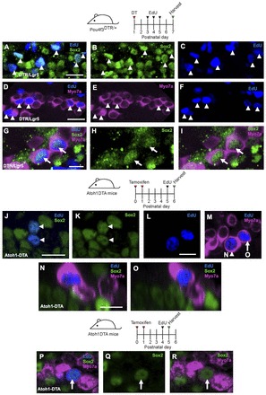
Fig. 5.

Mitotic HC regeneration in the neonatal mouse cochlea. Confocal images of EdU (blue) incorporation in Sox2+ SCs (green, A-C) and Myo7a+ cells (magenta, D-F) in the apical turn of Pou4f3DTR/+ mice at P7 after EdU injections at P3-P5. Some EdU+ HCs (Myo7a+) were also co-labeled with Sox2 (G-I). EdU+ SCs (Sox2, J,K) and EdU+/Myo7a+ cells (L,M) were also observed in the apical turn of Atoh1DTA mice 24 hours after EdU injection at P5. (N-O) Cross-section focused on the EdU+ nucleus of the cells indicated by the arrowhead and arrow in M. (P-R) Confocal images of EdU+ (blue) HCs (Myo7a, magenta) co-labeled with Sox2 (green) in the apical turn of Atoh1DTA mice 24 hours after EdU injection at P4. Scale bars: 20 μm in A-F; 10 μm in G-O.
