Fig. 6.

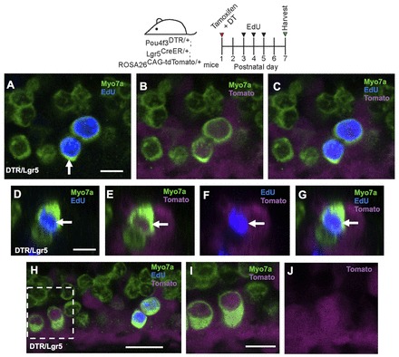
Both mitotic regeneration and direct transdifferentiation occur in the neonatal mouse cochlea. (A-C) Confocal images of tdTomato+ (magenta) HCs (Myo7a, green) that are labeled by EdU (blue) in the apical turn of Pou4f3DTR/+; Lgr5CreER/+; ROSA26CAG-tdTomato/+ mice at P7 after EdU injections at P3-P5. (D-G) Cross-section focused on the tdTomato+/EdU+ HC indicated by the arrow in A. Note that GFP expression from the Lgr5CreER/+ allele is much weaker than EdU labeling. In the apical turn of the same organs, there were also EdU-/Myo7a+/tdTomato+ cells (H-J). (I,J) Higher magnification of the boxed region in H. Scale bars: 20 μm.
