
Fig. 7.
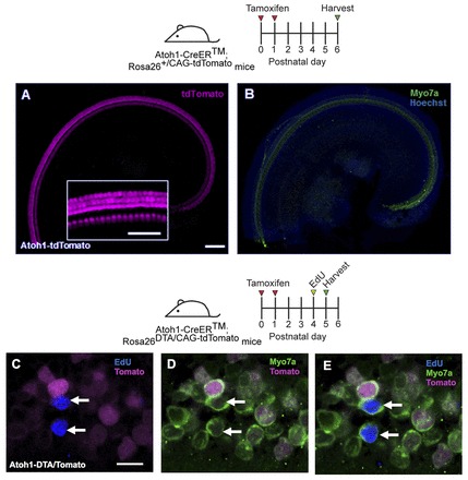
Regenerated HCs are not derived from original, differentiated HCs. (A,B) Confocal images of tdTomato+ (magenta) HCs (Myo7a, green) in the apical turn of Atoh1-CreER™; ROSA26CAG-tdTomato mice at P6 after tamoxifen injection at P0/P1. Nuclei are labeled by Hoechst (blue). Inset is a high-magnification image of tdTomato-labeled HCs. (C-E) Confocal images of EdU (blue) incorporation in Myo7a+ cells in the apical turn of Atoh1-CreER™; ROSA26DTA/CAG-tdTomato mice 24 hours after EdU injection at P4. DTA- HCs were traced with tdTomato (magenta). All EdU+/Myo7a+ cells were tdTomato-. Scale bars: 100 μm in A,B, 50 μm in inset; 10 μm in C-E.