Fig. 9.

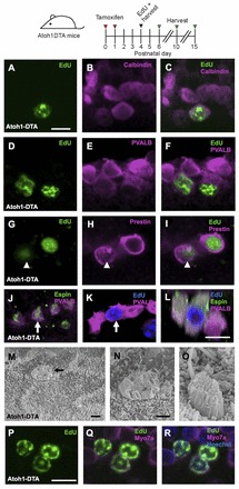
Regenerated HCs are similar to endogenous HCs. Confocal images of EdU+ (green) cells co-labeled with HC markers in the apical turn of Atoh1DTA mice. (A-C) Four hours after EdU injection at P5, EdU+ cells also express calbindin (magenta). (D-F) Two days after EdU injection at P4 (at P6), EdU+ cells express parvalbumin (PVALB; magenta). (G-I) Six days after EdU injection at P4 (at P10), EdU+ cells express prestin (magenta). Note that prestin is expressed in the cytoplasm of the EdU+ HC indicated by the arrowhead, which is characteristic of a young HC (Mahendrasingam et al., 2010). (J-L) Eleven days after EdU injection at P4 (at P15), EdU+ cells (blue) have espin+ (green) stereocilia bundles. (L) Cross-section focused on the EdU+ HCs indicated by the arrow in J and K. (M-O) Scanning electron micrographs of the apical turn in Atoh1DTA mice at P15. (N) High-magnification image of M, showing an immature stereocilia bundle. The immature bundle in O still has a kinocilium. (P-R) Confocal images of EdU+ (green) HCs (Myo7a, magenta) in the apical turn of Atoh1DTA mice at P10 after EdU injection at P4. Scale bars: 10 μm in A-L,P-R; 1 μm in M-O.
