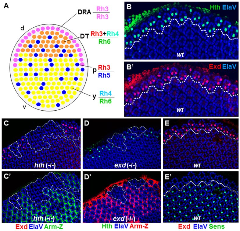
Fig. 1.

Extradenticle and Homothorax colocalize in the nuclei of DRA inner photoreceptors. (A) Schematic of the Drosophila retinal mosaic: a band of of specialized ommatidia with monochromatic R7 and R7 (Rh3) are always found in the DRA (pink). Dorsal third ommatidia are directly adjacent (orange); Rh3 and Rh4 are co-expressed in R7, and R8 express Rh6. Two additional subtypes are distributed randomly through the rest of the retina, named ‘pale’ (p) (Rh3/Rh5; shown in blue), and ‘yellow’ (y) (Rh4/Rh6, shown in green). (B) Colocalization of Hth (green) and Exd (red) in pupal DRA R7 and R8 nuclei (dashed line). Nuclear localization of Exd (B′) was observed in all Hth-expressing photoreceptors (B). (C) Exd (red) was lost in homozygous hthP2 (-/-) clones in the DRA (dashed lines), marked by absence of Armadillo-lacZ (green) (C′). (D) Hth (green) was lost in homozygous exd1 (-/-) clones at the DRA, marked by absence of Armadillo-lacZ (red, D′). (E) In wild-type pupae, the R8 marker Sens (green) was specifically excluded from R8 cells of DRA ommatidia (dashed white line), marked with Exd (red) (E′). d, dorsal; DT, dorsal third; v, ventral.
