
Figure 2.
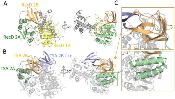
Structural homology between TSA and RecD helicase.A. Domain structure of RecD. The structure of a RecD homologue (RecD2 from Deinococcus radiodurans; PDB ID PD 1274) is shown in ribbon representation and in two orientations, 120° apart along the vertical axis. Domains 1A, 2A and 2B of RecD2 are colour-coded yellow, green and orange respectively.B. Superposition of the structure of TSA onto the structure of RecD2. RecD2 is in the same orientation as in (A) but is colour-coded in grey. TSA is in the same orientation and same colour-coding as in Fig. 1D. Orange and green rectangles indicate the regions, the details of which are shown in (C).C. Zoom-in on the superimposed parts. The regions within the orange and green rectangles in (B) are zoomed in to provide a visual illustration of the structural superposition between equivalent domains in TSA and RecD2. As can be seen, the 2A and 2B domains of TSA superimpose well onto parts of the 2A and 2B domains of RecD2. However, the 2B-like domain is shown not to be a part of the helicase domain.