
Figure 6.
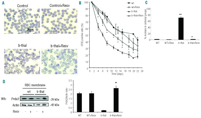
In vivo supplementation with resveratrol ameliorates red cell morphology, increases red cell lifespan and reduces red cell membrane oxidative damage in β-thalassemic mice. (A) Morphology of red cells from wild-type (wt) and β-thalassemic (b-thal) mice with or without resveratrol supplementation. Cells were stained with May-Grunwald-Giemsa. Cells were imaged under oil at 100× magnification using a Panfluor objective with 1.30 numeric aperture on a Nikon Eclipse DS-5M camera and processed with Digital Slide (DS-L1) Nikon. We show one representative image from a total of 12 for each condition. (B) Red cell survival (see also Methods) of CFSE labeled red cells from wild-type (wt) and β-thalassemic (b-thal) mice with or without resveratrol (Resv) supplementation. Data presented as means ± SD (n=4) from each mouse group; *P<0.05 compared to untreated mice. (C) Percentage of carbonyl groups from red cell membranes from wild-type (wt) and β-thalassemic (b-thal) mice with or without resveratrol (Resv) supplementation. Data are presented as means ± SD (n= 6) from each group; *P<0.05 compared to untreated mice; °P<0.05 compared to wild-type mice. (D) Peroxiredoxin-2 (Prdx2) membrane association in wild-type (wt) and β-thalassemic (b-thal) mice with or without resveratrol (Resv) supplementation. Actin was used as loading control protein. (Right): relative quantification of immunoreactivity of peroxiredoxin-2 (Prdx2) and actin in red cell membrane from wild-type (wt) and β-thalassemic (b-thal) mice with or without resveratrol (Resv) supplementation. Data are presented as means ±SD (n=6); *P<0.05 compared to untreated mice.