Figure 5.

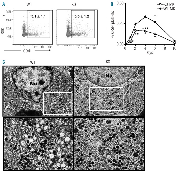
Functional and morphological abnormalities in KO megakaryocytes. (A) Representative FACS plots of CD41+ megakaryocytes prepared from 8 week-old WT and IEX-1 KO BM cells. (B) Insufficient production of platelets from IEX-1 KO megakaryocytes. CFSE-stained megakaryocytes of indicated genotypes were transferred into WT cognate mice at 5×10⁵ cells/mouse. The resultant platelet production was evaluated at indicated days and expressed as percentages ± SD of CFSE+ platelets relative to total platelets in circulation. *, **, and ***, P<0.05, 0.01 or 0.001, respectively, in the presence or absence of IEX-1. Data in A and B were collected from 2 independent experiments each with 5 mice per group. (C) Transmission electron microscopy (TEM) of megakaryocytes. BM cells were isolated from 8-week old WT and KO mice and subjected to TEM sample processing. Shown are the finely structured DMS of a mature WT megakaryocyte (left) and abnormal DMS of a mature KO megakaryocyte (right). Original magnification 6,000× in the upper panel, in which highlighted areas are enlarged in the bottom panels.
