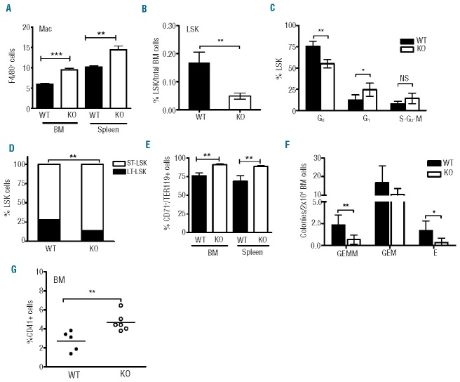
Figure 7.
LSK exhaustion and compromised progenitor function in IEX-1 KO mice after TBI. (A) An increase in the proportion of macrophages (MØ) marked as F4/80+ cells in the BM and spleen of IEX-1 KO mice compared to WT mice after eight months of TBI. (B) A marked decrease in the percentages of LSK cells in KO mice in comparison with WT mice after eight months of TBI as analyzed by flow cytometry as Figure 1A. (C) Increased cell cycling of KO LSK cells determined similarly as Figure 1B. (D) A decreased LSK pool in IEX-1 KO BM after TBI. Relative percentages of long- and short-term LSK cells as a total LSK cell population were analyzed in irradiated WT and KO BM on the basis of CD135 and CD34 expression as detailed in Methods. (E) Increased erythroblasts within the BM and spleen of irradiated KO mice, quantified as percentages ± SD of TER119+/CD71+ cells relative to a total TER119+ cells. (F) Colony forming assays of BM cells isolated from radiated KO (unfilled) and WT (filled) mice. Shown are the colony numbers of mixed multi-lineage (GEMM), granulocyte-monocyte (GM), and erythroid (E) progenitors per 2×10⁴ BM cells. N=6 for each culture condition. (G) IEX-1 KO megakaryocytes increase in number in KO BM compared to WT BM controls after TBI. Each symbol represents data from individual mice and horizontal lines indicate the mean in (G). *, ** and ***, P<0.05, 0.01 or 0.001, respectively, in the presence versus absence of IEX-1, and n= 12 for IEX-1 KO mice or 8 for WT mice for all studies except for colony forming cultures (F).

