Figure 8.
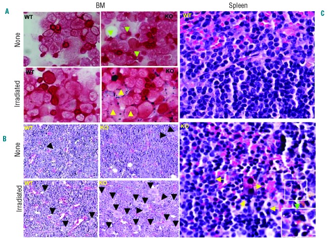
Histopathological examination of BM and spleens after TBI. BM brushings prepared from WT and KO mice without radiation (none) and eight months post-radiation (irradiated) were stained with an HT20 Iron staining kit (Sigma) (A). Ringed sideroblasts were seen only in irradiated KO BM (yellow triangles, bottom right) and coarse iron deposits in non-irradiated KO BM (green triangles, upper right) in (A). Moreover, internuclear bridges (yellow arrows) and binucleated erythroid precursors (white stars) were readily recognized in H&E staining of the spleen prepared from KO mice compared to WT spleens in eight months post-TBI (C). One of the internuclear bridges is enlarged in the middle and binucleated erythroid precursors in the white rectangle are magnified in the lower right corner in C (green arrow). H&E staining of sections prepared from WT and IEX-1 KO BM showed increased megakaryocytes in irradiated IEX-1 KO mice (right bottom) compared to non-irradiated mice (right upper) or non-irradiated or irradiated WT controls (left panels) (B). Megakaryocytes are marked by filled triangles and original magnification 20X.

