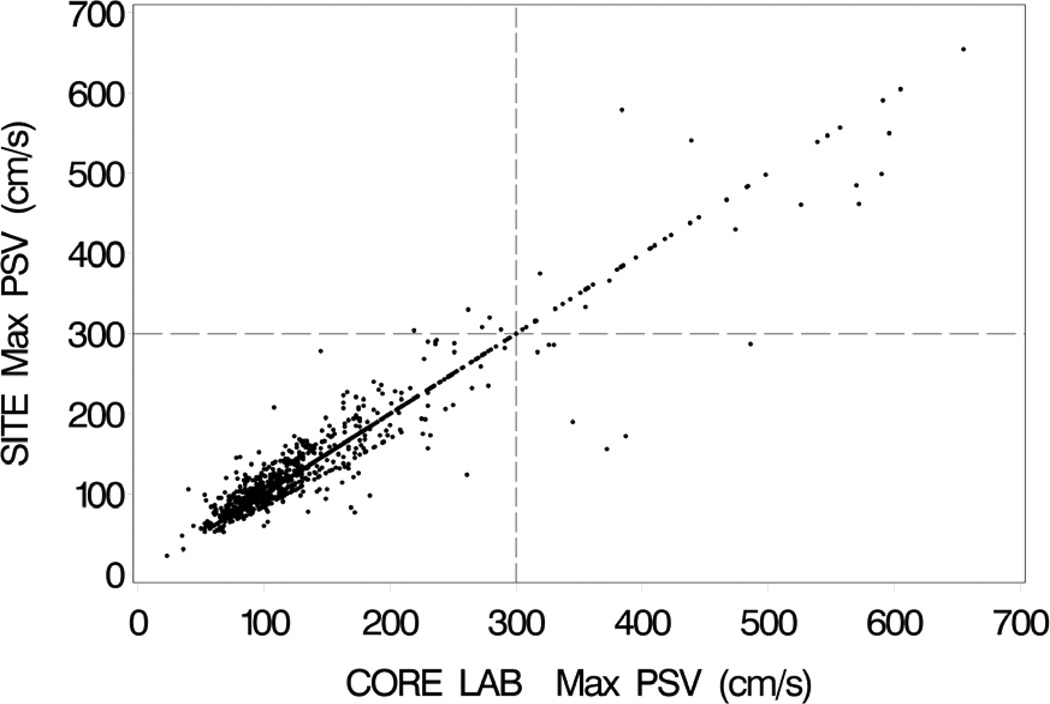
Figure 2.
Clinical site (SITE) maximum peak systolic velocities (PSV) on the vertical axis vs. UCL-verified (CORE LAB) maximum peak systolic velocities on the horizontal axis for the 12-month follow-up duplex scans. Both carotid stent and carotid endarterectomy cases are included. Vertical and horizontal dashed lines mark the velocity threshold of 300 cm/s. The concentration of data points along the “line of unity” represents exact agreements between the clinical sites and the UCL.

