Fig. 3.
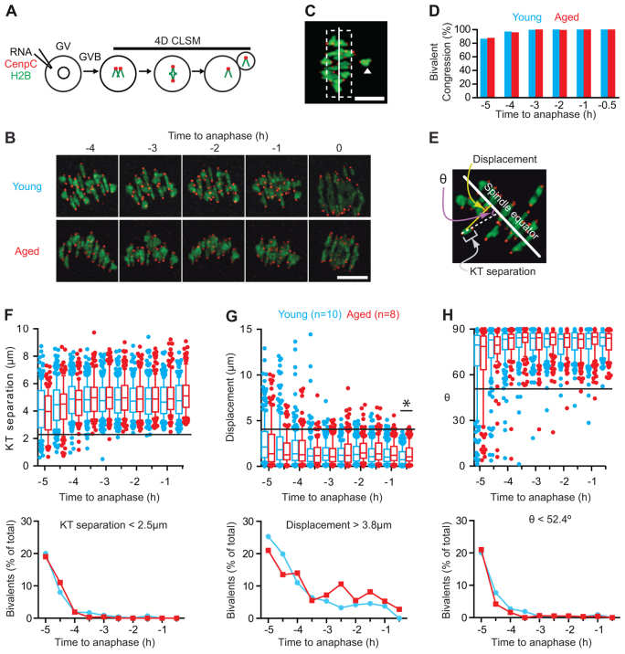
Congression and establishment of tension for bivalents are not affected by age. (A,B) Method used for chromatin and kinetochore labelling in live oocytes (A), and the resulting bivalent images at selected time points in young and aged oocytes (B). (C) A meiotic spindle, with one bivalent (arrowhead), classified as nonaligned because it is >4 μm (dashed line) from the spindle equator (solid line). (D) Percentage of congressed bivalents in young (n=10) and aged (n=8) oocytes at the times indicated. (E-H) Three parameters used on bivalents and their kinetochores (E); the separation distance between the two pairs of sister kinetochores (F, top), the displacement of the midpoint of the bivalent from the equator (G, top), and the bivalent axis of orientation (θ) relative to the spindle equator (H, top). Horizontal lines indicate minimum (F,H) or maximum (G) measurements made on young oocytes at the -0.5 hour time point; bivalents outside the lines were considered ‘at risk’ of mis-segregation. The percentage of bivalents found to be ‘at risk’ for each time point (F-H, bottom). Box plots, whiskers at 10-90 centiles; the asterisk indicates P<0.05 (Kruskal-Wallis). Scale bar: 10 μm in B,C.

