
Figure 5.
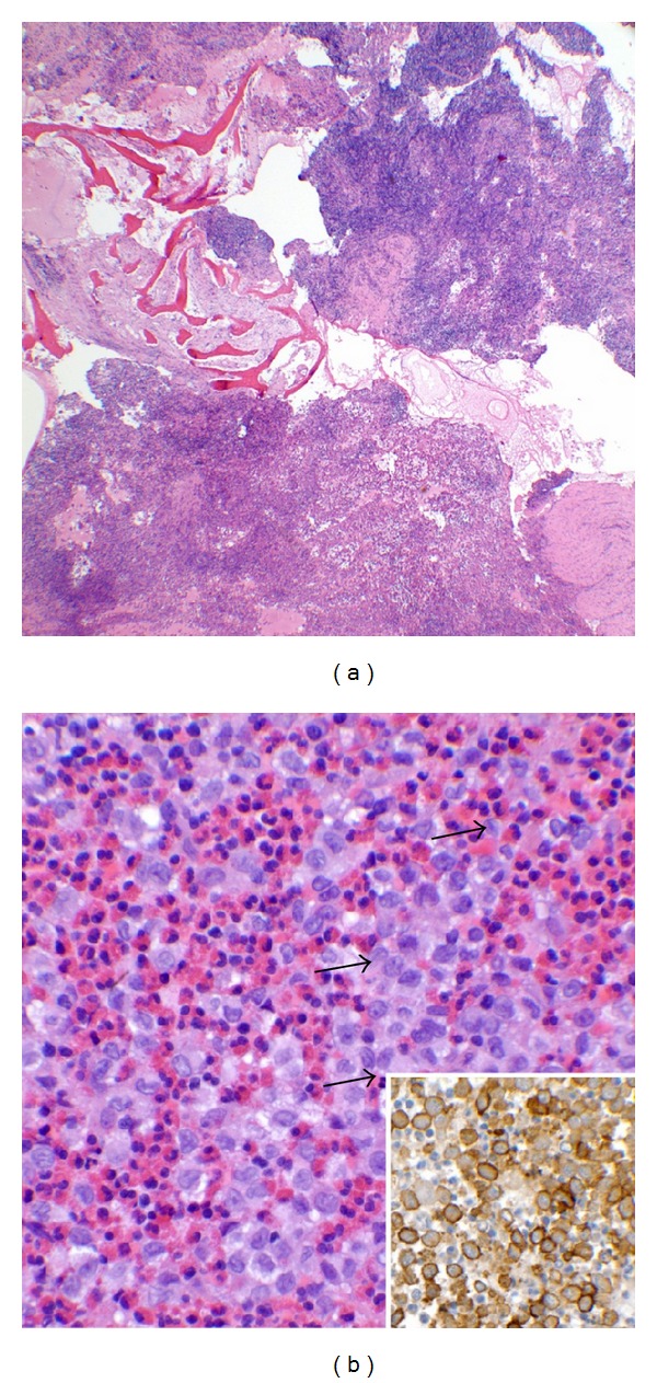
(a) Low magnification shows tumor cells expanding and destroying bony trabeculae (hematoxylin-eosin stain, original magnification ×40). (b) High magnification demonstrates tumor cells characterized by coffee bean-shaped grooved nuclei indicated by the arrow, admixed with eosinophils consistent with Langerhans cells (hematoxylin-eosin, original magnification ×400). Insert: CD1a immunostain is a transmembrane antigen normally found on Langerhasn cells (CD1a immunostain, original magnification ×600).