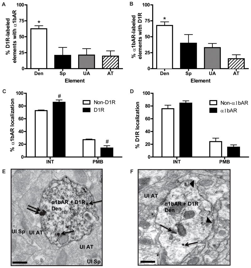
Fig. 5. Co-localization and distribution of D1R and α1bAR in the PFC.
(A) Mean ± SEM percent of D1R immunoperoxidase-labeled elements that also contain α1bAR immunogold labeling. N=4 rats. Total number of D1R-labeled elements examined: 91 dendrites, 12 spines, 35 unmyelinated axons, 38 axon terminals. *p<0.05 compared to each other type of element. (B) Mean ± SEM percent of α1bAR immunoperoxidase-labeled elements that also contain D1R immunogold labeling. N=5 rats. Total number of α1bAR labeled elements examined: 211 dendrites, 43 spines, 166 unmyelinated axons, and 89 axon terminals. *p<0.05 compared to each other type of element. (C) Mean ± SEM percent of INT and PMB α1bAR immunogold particles in dendrites that contain (D1R) or do not contain (non-D1R) D1R immunoperoxidase labeling. #p<0.05 compared to non-D1R for that location. (D) Mean ± SEM percent of INT and PMB D1R immunogold particles in dendrites that contain (α1bAR) or do not contain (non-α1bAR) α1bAR immunoreactivity. (E) Representative electron micrograph of a double labeled dendrite (Den) for D1R (immunoperoxidase, diffuse throughout) and α1bAR (immunogold). An unlabeled spine (Ul Sp) and axon terminal (Ul AT) are also indicated. (F) Representative micrograph of a dendrite with α1bAR immunoperoxidase labeling (arrowheads) and D1R immunogold labeling. For (E) and (F), single arrows indicate intracellular immunogold labeling, while double arrows indicate extrasynaptic plasma membrane bound labeling. Scale bars = 0.5 μm.

