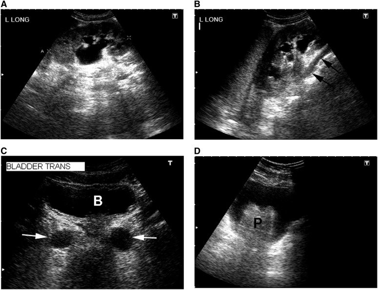
Figure 2.
Sonographic appearance of the upper and lower urinary tract. (A) Hydronephrosis of a left kidney (longitudinal view) with dilated renal pelvis and major and minor calyces. (B) Another longitudinal view of the same kidney showing a dilated ureter (arrows) tracking underneath the lower pole. (C) Transverse view of the urinary bladder (B) showing dilated distal ureters (arrows). (D) Transverse view of the urinary bladder with an enlarged prostate gland (P).

