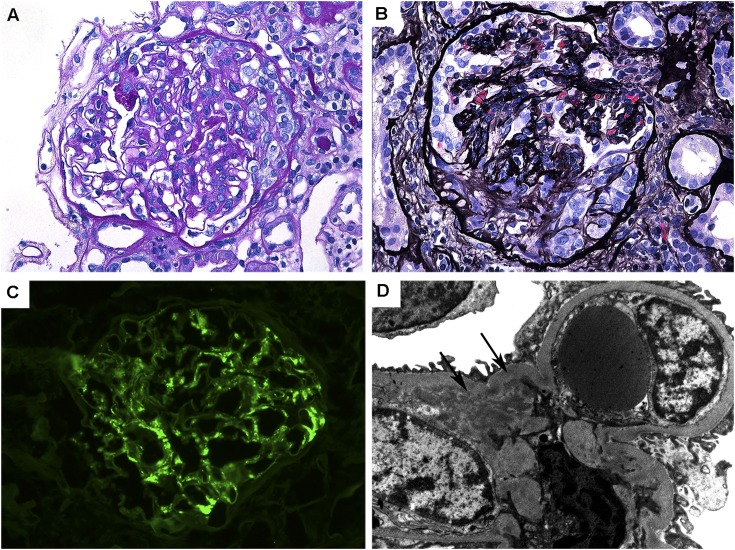
Figure 1.
Pathologic features of IgA nephropathy associated with inflammatory bowel disease. (A) Glomerulus with mild segmental mesangial matrix expansion and mesangial hypercellularity (periodic acid-Schiff, original magnification ×400). (B) Glomerulus with a small cellular crescent and compression of the underlying glomerular tuft (Jones methenamine silver, original magnification ×400). (C) Glomerulus with global mesangial staining with antisera for IgA (fluorescein, original magnification ×400). (D) Electron microscopy of mesangial region containing small electron-dense deposits (arrows) (original magnification ×12,000).

