
Figure 7.
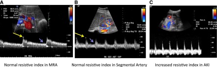
Doppler imaging and resistive index. (A) Normal color and spectral Doppler ultrasonogram of the right main renal artery (MRA) demonstrates normal peak systolic velocity (98.5 cm/sec), resistive index (0.67), and acceleration time (32 milliseconds). (B) Normal color and spectral Doppler ultrasonogram of the right superior segmental renal artery (SEG ART SUP) demonstrates normal resistive index (0.68). (C) Spectral Doppler image of left inferior segmental renal artery demonstrates an abnormally high resistive index of 0.76 in a 39-year-old woman with multiple myeloma and AKI. Resistive index is determined by the following formula: (peak systolic velocity − end diastolic velocity)/(peak systolic velocity); in A and B, peak systolic velocity is indicated by the long yellow arrows, and end diastolic velocity is indicated by the short purple arrows. AO, aorta.