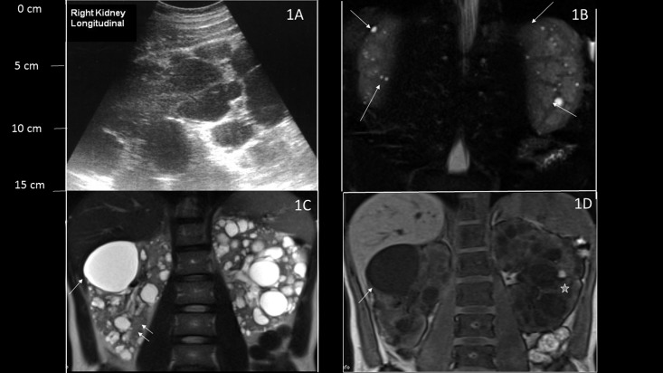
Figure 1.
Ultrasonography and magnetic resonance imaging (MRI) imaging of patients with autosomal dominant polycystic kidney disease (ADPKD) compared with bilateral simple acquired cysts. (A) Longitudinal ultrasonographic view of the right kidney showing multiple dark hypoechoic lesions throughout the kidney with kidney enlargement, consistent with ADPKD. (B) Coronal T2-weighted MRI images show multiple tiny T2 high signal lesions in both kidneys with well preserved renal parenchyma in a patient with bilateral acquired cysts (arrows). (C) Coronal T2-weighted and (D) coronal unenhanced three-dimensional T1-weighted gradient echo MRIs of a patient with ADPKD and conserved renal function show multiple cystic lesions. Lesions with high T2 signal and low T1 signal represent simple cysts (arrow), whereas hemorrhagic cysts show low T2 signal and high T1 signal (star). Preserved renal parenchyma is seen between the cysts (double arrow).

