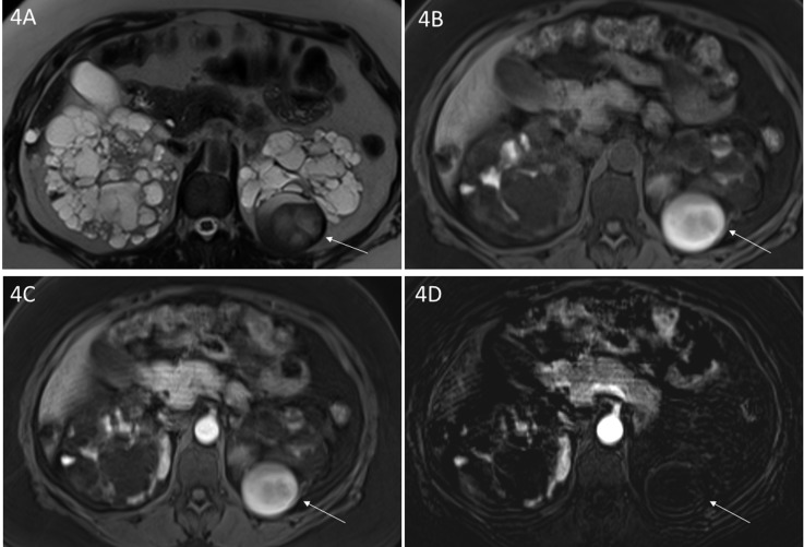
Figure 4.
Complex cyst in a patient with autosomal dominant polycystic kidney disease. (A) Axial T2-weighted and axial (B) unenhanced and (C) enhanced three-dimensional T1-weighted gradient echo magnetic resonance images depict a mass in the lower pole of the left kidney with (A, arrow) heterogeneous T2 signal intensity and (B, arrow) T1 high signal. After administration of Gadobenate Dimeglumine, there is high signal on (C, arrow) an enhanced image similar to the (B) unenhanced T1-weighted image. The absence of enhancement on the (D, arrow) subtraction image supports the diagnosis of complex hemorrhagic cyst and rules out a tumor.

