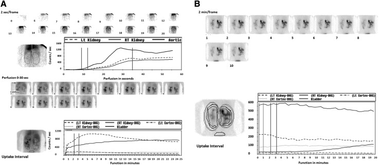
Figure 5.
Mercaptoacetyltriglycerine-3 (MAG-3) renal scintigraphy without and with injection of furosemide in a 33-year-old patient with autosomal dominant polycystic kidney disease who presented with massive gross hematuria and acute renal failure. A, top, represents the perfusion phase, and the bottom images show the excretion phase before injection of furosemide. (B) The postfurosemide images show asymmetric excretion and a higher intensity signal on the right side. The times to peak height of the cortical renogram curves are 2.72 for the left kidney and 16 for the right kidney. The cortical 20 minutes to maximum ratios are 0.48 for the left kidney and 0.98 for the right kidney. These results suggest obstruction on the right side, which in this case, was caused by ureteral clots.

