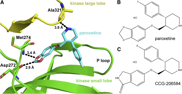
Fig. 1.
Structure of paroxetine bound to GRK2 and of inhibitors used in this study. (A) Paroxetine in the active site of GRK2 in the GRK2·paroxetine–Gβγ complex (PDB ID 3V5W). The kinase domain large lobe is colored yellow and the small lobe green. Paroxetine is drawn as a stick model with carbons colored cyan, oxygens red, and nitrogens blue. Three hydrogen bonds, including an unconventional C–O hydrogen bond, are shown as black dashed lines with associated distances. Chemical structures of paroxetine (B) and its benzolactam derivative CCG-206584 (C) are shown.

