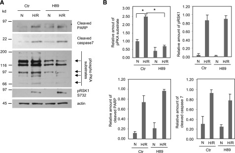
Fig. 6.

Elevated PKA activity does not contribute toward the H/R-induced apoptosis in cardiomyocytes. (A) After serum starvation, HL-1 cells were treated with or without H89 (10 μM) and exposed to normoxia or hypoxia (1% O2) for 24 hours, followed by reoxygenation for 1 hour. Cell lysates were subjected to Western analysis for the indicated proteins. Arrows depict the changes in intensity of the PKA substrate bands. (B) Quantitative data from Western blots of three different experiments normalized for actin (loading control). To quantify the amount of phospho-PKA substrates, the 66-kDa PKA substrate bands were scanned and normalized for actin. Data shown are means ± S.E.M. *P < 0.05. Ctr, control; N, normoxia.
