
Figure 3. Effects of MSC seeding and MSC sheets on cartilage and bone formation at 4- and 6-weeks post-surgery.
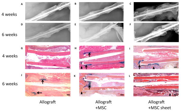
The upper panels (A, B, C, D, E, F) are representative x-ray images of allografts, allografts seeded with MSCs, and allografts wrapped with MSC sheets at 4- and 6-weeks post-surgery. Lower panels (G, H, I, J, K, L) show the histological sections stained with AB/H/OG. (Scale bars=400μm). Bone callus is marked with black triangles and cartilaginous callus is marked with black arrows.