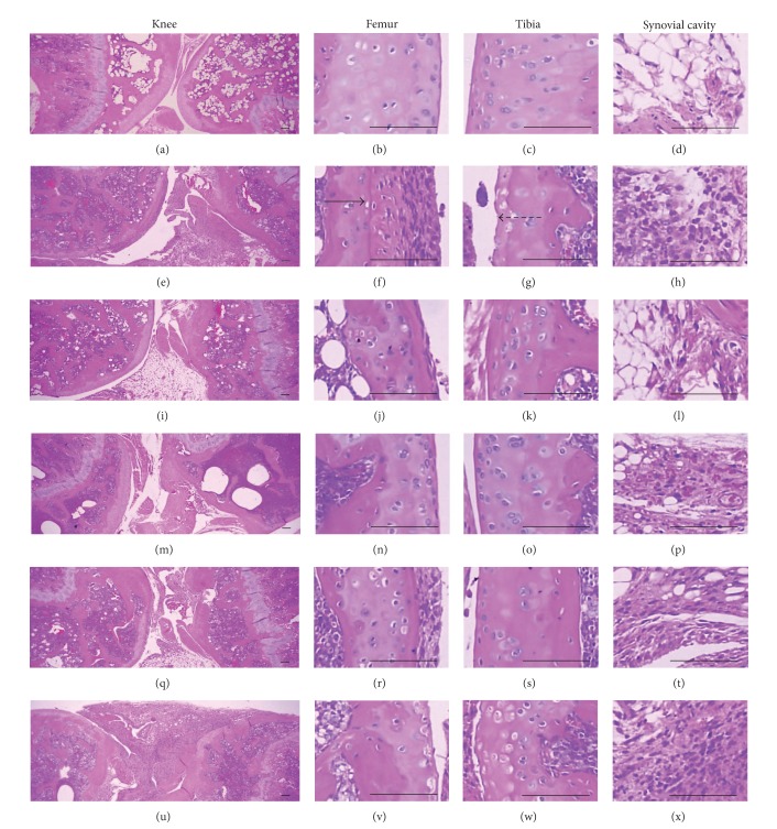
Figure 5.
Histopathological profiles of the knee in the intact control ((a)–(d)), CIA control ((e)–(h)), Enbrel group ((i)–(l)), platycodin D-treated groups; 200 mg/kg ((m)–(p)), 100 mg/kg ((q)–(t)), and 50 mg/kg ((u)–(x)). Marked decreases in the articular surfaces (cartilage and bone) were detected in the knee articular surfaces of the femur and tibia, with severe inflammatory cell infiltration into the synovial cavity in the CIA control mice. However, histopathological changes in the CIA group were decreased dramatically by treatment with platycodin D. The arrow indicates articular surface thickness. Dotted arrow indicates articular cartilage thickness. All were stained with H&E. Scale bars = 160 μm.

