
Figure 7.
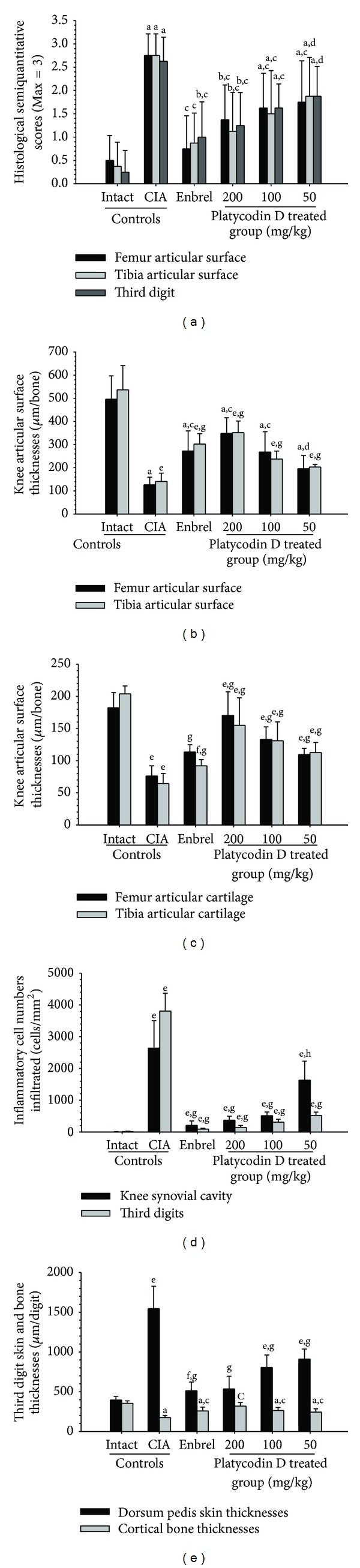
Platycodin D ameliorated the histopathological changes of the knee and third digit. (a) The semiquantitative scores of platycodin D-treated mice were lower compared with the CIA control mice. (b) The knee articular surface thickness of platycodin D-treated mice was greater compared with the CIA control mice. Knee articular surface thicknesses are shown in Figure 5 (arrow). (c) The knee articular cartilage thickness of platycodin D-treated mice was greater compared with the CIA control mice. Knee articular cartilage thicknesses are shown in Figure 5 (dotted arrow). (d) Infiltrated inflammatory cells of platycodin D-treated mice were lower compared with the CIA control mice (e). Increases in the third digit dorsum pedis skin thickness and decreases in the cortical bone thickness were inhibited by treatment with platycodin D compared with the CIA control mice. Measurements of the third digit dorsum pedis skin (arrow) and cortical bone (dotted arrow) thicknesses are shown in Figure 6. Values are expressed as means ± SD (n = 8); a P < 0.01 and b P < 0.05 as compared with intact control by LSD test; c P < 0.01 and d P < 0.05 as compared with CIA control by LSD test; e P < 0.01 and f P < 0.05 as compared with intact control by MW test; g P < 0.01 and h P < 0.05 as compared with CIA control by MW test.