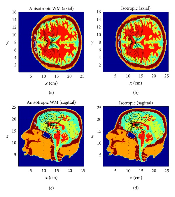
Figure 7.

Impact of brain white matter (the red tissue) anisotropy on the EEG forward solution: axial views (top); sagittal views (bottom). The white matter is anisotropic (left) and isotropic (right). A horizontally oriented dipole is placed deep in the central brain region. The distortion of current stream lines can be seen in the anisotropic case (left) relative to the isotropic case (right).
