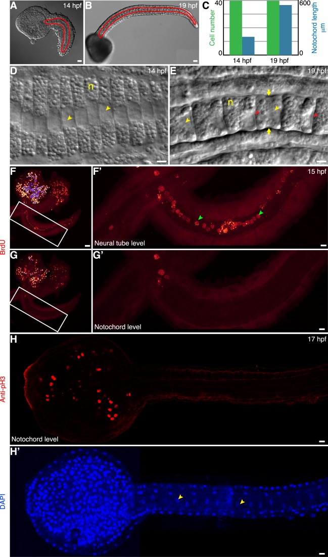
Figure 1. Notochord cells elongate and are postmitotic.
(A) A Ciona embryo at 14 h postfertilization (hpf). (B) A Ciona embryo at 19 hpf. The notochord, outlined in red, is located centrally in the tail. (C) Between 14 and 19 hpf, the notochord cell number remains 40, whereas the notochord length increases by approximately 2.5-fold. (D) Notochord cells (n) at 14 hpf are coin shaped. (E) At 19 hpf, these cells have become drum shaped. A circumferential constriction (yellow arrow) is present at the halfway between the two ends of the cylindrical cell and persists during elongation. Yellow arrowhead indicates the nucleus, which is positioned at the posterior end of the cell. Red arrowhead indicates the lumen emerging after 18 hpf. (F, enlarged in F′) BrdU labeling of cells in the head and the tail at the level of dorsal neural tube at 15 hpf. Green arrowheads point to BrdU-positive nuclei of the cells in the neural tube. (G, enlarged in G′) A section through the notochord of the same embryo shows the absence of BrdU incorporation in the notochord cells. (H and H′) Confocal section at the level of the notochord of a 17 hpf embryo labeled with anti-pH3 and counterstained with DAPI. Anti-pH3 labels cells in the head, but not in cells of notochord, whose nuclei are visualized by DAPI (H′, indicated by yellow arrowheads). Scale bar in (D, E, F′, G′, H, H′), 5 µm; in (A, B, F, G), 20 µm. Anterior to the left.

