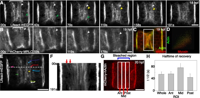
Figure 4. Cortical flow contributes to the formation of the equatorial actomyosin ring.
(A) Time-lapse projections of a notochord cell showing that lifeact-mEGFP–labeled actin filaments (white and yellow arrowheads follow two circumferential actin filaments) move toward and align parallel to the equator in the equatorial region (bracket) (see also Movie S3). In addition to the circumferential long actin filaments, between the lateral domain and the equatorial region, a population of longitudinally oriented short filaments (green arrowhead follows a single filament) move into the equatorial region, where they join the circumferential filaments. (B) Dynamic movement of mCherry-MRLC–labeled myosin filaments toward the equator in the equatorial region (bracket). White arrowhead follows a myosin filament. (C) Confocal section (1 µm) of a notochord cell double labeled for actin (lifeact-mEGFP) and myosin (mCherry-MRLC) shows that actin filaments and myosin filaments are present simultaneously in the contractile ring. (D) Colocalization analysis of the equatorial region (box in C) shows that a significant amount of actin filaments and myosin filaments do not colocalize. (E) Manual tracking of single actin filament movement (color-coded) in a notochord cell (see Movie S3). The average velocity is 33.9±4.9 nm/s (mean ± s.e.m., n = 9). (F) Kymograph of actin filament movement (indicated by arrows) based on Movie S3 at the location indicated by the dash line in (E). The velocity is 29.5±10.5 nm/s, n = 2. (G and H) Dynamics of actin filament movement revealed by FRAP. (G) A projection of a mCherry-hActin–expressing cell that was photobleached. The photobleach mark was made over the entire equatorial region, and the halftime for recovery was determined in both the whole region, and separately in the anterior (ant), middle (mid), and posterior (post) regions, and is shown in (H). Recovery is faster in the anterior and posterior regions than in the middle region. This confirms that actin filaments flow toward the equator. Actin movement velocity is 40.8±3.8 nm/s, n = 4, calculated from the recovery kinetics of the whole region. Scale bars, 5 µm.

