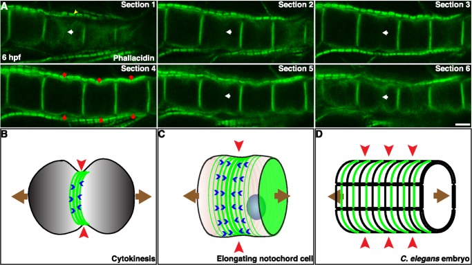
Figure 7. Conservation of equatorial actin ring and circumferential contraction as a common and mechanistically scalable biophysical solution for anisotropic shape change.
(A) Confocal sections of elongating notochord cells in an appendicularian Oikopleura dioica embryo stained with phallacidin show the presence of an equatorial constriction (red arrows) and actin ring (white arrows). Yellow arrowhead indicates the actin filaments in muscle sarcomeres. (B) The equatorial circumferential constriction (red arrowhead) created by an actomyosin ring (green lines) in a dividing cell causes cell elongation (brown arrows) before cell division. Blue arrowheads indicate filament flow. (C) In notochord cells, an actomyosin ring (green lines) causes an equatorial constriction (red arrowhead), which contributes to the cell elongation (brown arrows). (D) During C. elegans embryogenesis, numerous circumferential actin filaments (green lines) are present at the outer surface of the hypodermal cells. The elongation of the embryo (brown arrows) is accomplished by the contractility of these actin filaments squeezing the embryo circumferentially (red arrowheads) at the scale of the entire embryo. Scale bar, 5 µm.

