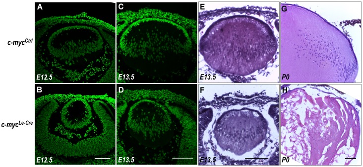
Figure 2. Defects in lens embryonic development of c-myc-deficient lens.
(A–D) Representative confocal pictures of E12.5 (A–B) and E13.5 (C–D) control (c-mycCtrl) and c-myc-deficient (c-mycLe-Cre) lens cryosections stained with sytox green. The pattern of nuclear staining of c-myc–deficient and control lens indicates a developmental delay. (E–H) Representative pictures of E13.5 sections of c-mycLe-Cre and c-mycCtrl lens stained with hematoxilin and eosin (E–F) and P0 (G–H). At P0, the morphology of c-myc-null lens is severely compromised. Scale bar: 100 μm.

