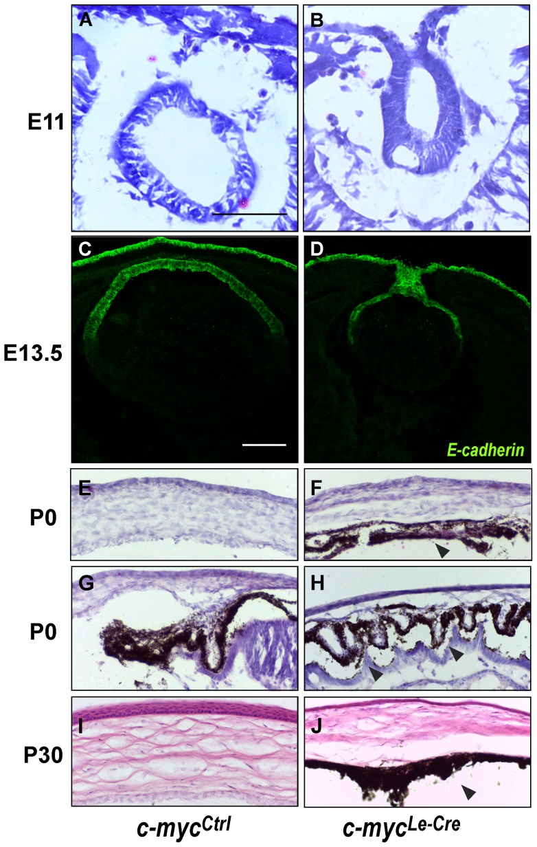
Figure 3. Loss of c-myc leads to development defects in the anterior chamber.
Representative pictures of hematoxylin & eosin (H&E) staining at E11.0 (A, B) and immunofluorescence for E-cadherin at E13.5 (C, D) illustrates the defective lens vesicle formation, which leads to the formation of the lens stalk. (E–J) H&E staining of anterior segment at P0 (E–H) and P30 (I–J) demonstrates that c-mycLe-Cre eyes presented corneal stroma loosening, absence of corneal endothelium and presence of pigmented cells along the anterior chamber (at P0 and P30, arrowheads in F, H and J). Scale bar: 100 μm.

