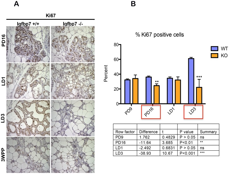
Figure 6. Igfbp7−/− glands show decreased proliferation index.
(A) Inguinal mammary glands were isolated from wild-type (Igfbp7+/+) or the Igfbp7 −/− mice on pregnancy days 9 (PD9) and 16 (PD16) as well as on lactation days 1 (LD1) and 3 (LD3) and at 3 weeks postpartum (3WPP). Sections from the formalin fixed and paraffin embedded glands were used to detect the expression of Ki-67 protein by Immunohistochemical stains. As can be seen, the Igfbp7 −/− glands show decreased number of cells positive for the expression of Ki-67. (B) The proliferation index of the WT and the Igfbp7 −/− glands are determined by examining the nuclear expression of Ki-67 protein in over 600 nuclei in the slide sections. The proliferation index is calculated by obtaining the percentage of Ki-67 nuclei positive cells in each section. The average values obtained from 3 different fields and the standard deviations are plotted again the developmental stage of the mammary glands. P-values were obtained by performing two-tailed T-tests and provided in a table under the graph.

