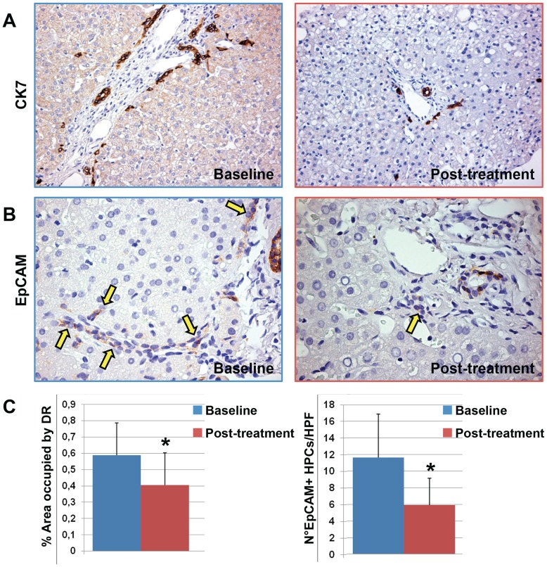
Figure 1. Immunohistochemistry for Cytokeratin(CK) 7 and EpCAM in liver biopsies of pediatric NAFLD patients.
A) at the beasline, pediatric NAFLD biopsies were characterized by a prominent expansion of hepatic progenitor cell (HPC) pool and the presence of reactive ductules at the periphery of portal spaces. After DHA treatment, a minimal involvement of the HPC compartment was present. Original magnification 20×. B) EpCAM-positive HPCs (arrows) were significant reduced by DHA treatment in comparison with the biopsies at the baseline. Original magnification 40×. C) Histograms show the reduction of HPC expansion (ductular reaction extension) and activation (number of EpCAM+ cells) after DHA treatment. Data are shown as Means ± Standard Deviation. * = p<0.05.

